

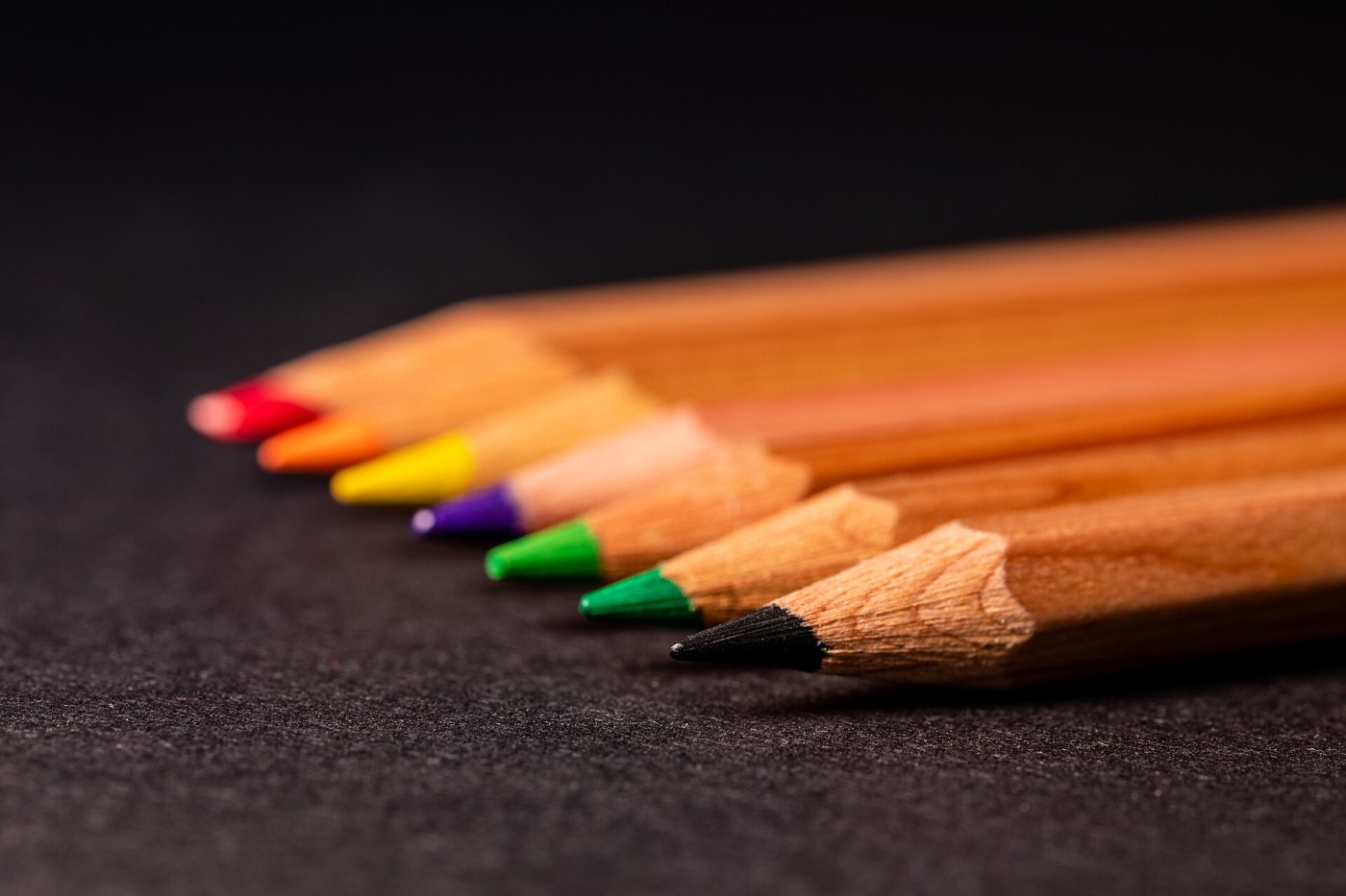
Hold a ripe strawberry next to its leaves and the red seems to belong to the fruit itself. Yet the light bouncing off it carries no colour at all: it is simply a mixture of electromagnetic waves of different lengths. As Isaac Newton noted in his 1704 book Opticks, light rays are not coloured in themselves. The red you experience is built inside your head, through a chain of events that starts with three kinds of light-sensitive cells in the eye and ends in specialised patches of the brain. That chain explains why about 1 in 12 men confuse certain colours, and why a photograph of a dress once split the internet in two.
Three cones and a sliver of the spectrum
Our eyes respond to only a narrow band of electromagnetic radiation, with wavelengths from roughly 380 to 700 nanometres (billionths of a metre). This light is captured by the retina, the thin layer of nerve tissue lining the back of the eye, which contains two main kinds of light detectors, or photoreceptors. Around 90 to 120 million rods work in dim light but cannot tell colours apart, which is why a moonlit garden looks grey. About 5 to 6 million cones work in daylight and make colour vision possible. They are packed most tightly in the fovea, the small central pit we aim at whatever we want to see sharply.
Cones come in three types, each carrying a slightly different light-absorbing protein called an opsin. Short-wavelength (S) cones respond best to violet-blue light around 420 nm, medium-wavelength (M) cones to green light around 530 nm, and long-wavelength (L) cones to yellowish-green light around 560 nm. They are nicknamed "blue", "green" and "red" cones, but their sensitivities overlap widely, and a single cone cannot tell a better-matched wavelength from simply brighter light. Colour appears only when the nervous system compares the signals from different cone types.
A 200-year-old idea, confirmed
In 1802 the English physician Thomas Young suggested that the eye needs only three kinds of sensors to produce every colour we perceive, and in the 1850s the German scientist Hermann von Helmholtz developed this into the trichromatic theory. Direct proof came in 1964, when two American teams, one including Edward MacNichol and the other led by George Wald, shone tiny beams of light through single cones from primate and human retinas and found three distinct absorption peaks. In 1986 Jeremy Nathans and colleagues at Stanford University identified the genes for the three cone opsins. The S-opsin gene lies on chromosome 7, while the L and M genes sit side by side on the X chromosome. Their proteins are about 96% identical, a sign that one ancestral gene was duplicated tens of millions of years ago in the ancestors of Old World monkeys, apes and humans.
When a cone is missing or shifted
Because the L and M genes are on the X chromosome, red-green colour vision deficiency is far more common in males, who have only one X. According to the US National Eye Institute, about 1 in 12 men have some form of it; in women it is much rarer, roughly 1 in 200. Some affected people lack one cone type entirely (dichromacy), and more have a cone whose sensitivity is shifted towards its neighbour (anomalous trichromacy), so that reds, browns, oranges and greens are hard to tell apart. Seeing no colour at all is very rare.
The first scientific description came from the English chemist John Dalton, who reported in 1794 that a flower others called pink looked sky blue to him in daylight. He guessed that the fluid in his eyes was tinted blue; when they were examined after his death, no tint was found. In 1995, researchers in Britain extracted DNA from his preserved eye tissue and showed that he lacked a working M-opsin gene: Dalton was a deuteranope, missing his medium-wavelength cones.
In animals, such a deficiency has even been reversed. In 2009 a team led by Jay and Maureen Neitz used a harmless virus to deliver a human L-opsin gene into the retinas of adult male squirrel monkeys born red-green colour blind. About 20 weeks later the monkeys began passing colour tests they had always failed. No such treatment is approved for people. Anyone who suspects a problem with their colour vision should see an eye doctor, who can test it with simple charts.
Opposing pairs: the retina compares
In the 1870s the German physiologist Ewald Hering pointed out that we can picture a reddish yellow (orange) or a bluish green (turquoise), but never a reddish green or a yellowish blue. He proposed that colour is coded in opposing pairs: red against green, blue against yellow, and light against dark. This opponent-process theory also explains afterimages: stare at a red square for half a minute, then look at a white wall, and a faint green square appears.
Both theories proved right, at different stages: the cones capture the light, and nerve cells in the retina then subtract their signals from one another. One channel compares L with M cones, producing a red-green signal; another compares S cones with the combined L and M signal, producing a blue-yellow signal; a third adds everything up to report brightness. In the 1960s Russell De Valois and colleagues recorded such colour-opponent cells in monkeys, in the lateral geniculate nucleus, the thalamus relay station between the eyes and the cortex.
Colour in the brain
From the thalamus, signals reach the primary visual cortex at the back of the head, then travel forward along the brain's underside. In 1973 Semir Zeki of University College London reported that many cells in an area of the macaque cortex now known as V4 respond selectively to colour. Brain imaging later revealed similar regions in people, on the underside of the occipital and temporal lobes. Damage there, usually from a stroke, can cause cerebral achromatopsia: the eyes work normally, yet the world looks like an old black-and-white film.
The brain also corrects for the lighting. White paper reflects orange-tinted light under a lamp and bluish light in shade, yet looks white in both. This colour constancy means the brain constantly estimates the colour of the light and discounts it. In 2015 a photograph of a striped dress showed what happens when the lighting is ambiguous: millions of people saw it as white and gold, while millions of others saw it as blue and black, which it really was.
Other eyes, open questions
Most mammals, including dogs and cats, have only two cone types, while many birds, reptiles and fish have four, including one tuned to ultraviolet or violet light. Some women carry two slightly different versions of the L or M gene and may have four cone types too, but in a 2010 study led by Gabriele Jordan at Newcastle University only one of 24 such women behaved as a true tetrachromat in demanding colour-matching tests. An extra cone does not automatically bring an extra dimension of colour.
Big questions remain. Researchers still debate how much colour processing happens in areas such as V4 rather than across the wider visual brain. Language may play a part too: a 2007 study found that Russian speakers, who have separate basic words for light blue (goluboy) and dark blue (siniy), were slightly faster at telling apart blues on opposite sides of that boundary. And no experiment can yet show whether your red feels the same as someone else's. What is clear is that colour is not waiting inside objects. It is a comparison, made by three kinds of cells and a brain that is constantly interpreting the light around us.
Sources: Science (1986), Nathans, Thomas & Hogness: Molecular genetics of human color vision: the genes encoding blue, green, and red pigments; Science (1995), Hunt et al.: The chemistry of John Dalton's color blindness; Nature (2009), Mancuso et al.: Gene therapy for red-green colour blindness in adult primates; Journal of Vision (2010), Jordan et al.: The dimensionality of color vision in carriers of anomalous trichromacy; US National Eye Institute (NIH): Color Blindness. Image: Dietmar Rabich / Wikimedia Commons (CC BY-SA 4.0).


Comments
Please login to leave a comment Sign in