

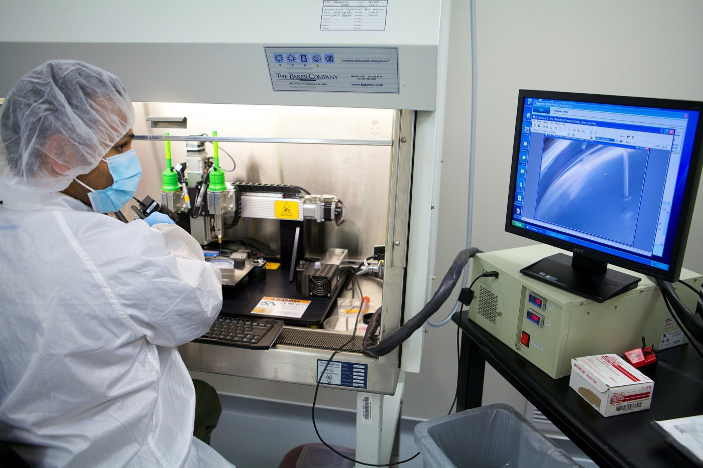
Inside a sterile cabinet, a nozzle glides back and forth over a glass dish, laying down a thin line of pale gel. Each pass adds another layer, thinner than a human hair. The machine looks much like the 3D printers that make plastic toys and spare parts, but the "ink" here is alive: a jelly packed with millions of human cells. When the printing is finished, the object is carried to an incubator, where the cells will begin to grow, attach to each other and, researchers hope, behave like real tissue. This is 3D bioprinting, one of the most ambitious ideas in modern medicine.
From inkjet cartridges to living ink
Ordinary 3D printing builds an object layer by layer from a digital design. Bioprinting applies the same principle to biology. The design often comes from a patient's own medical scans, such as CT or MRI images, so the printed shape can match a specific nose, ear or bone defect.
One of the early milestones came in the early 2000s, when Thomas Boland and colleagues at Clemson University in the United States modified ordinary desktop inkjet printers so that they could deposit proteins and living cells instead of ink. They found that many cells survived the trip through the tiny print head. Since then, the field has grown into a large international research effort, with university laboratories and companies building specialised machines.
The key ingredient is the bioink: a mixture of living cells and a supporting material, usually a hydrogel, a water-rich gel similar in texture to soft jelly. Common hydrogels include alginate from seaweed, gelatin and collagen, the main structural protein of the body. The gel must be runny enough to pass through a nozzle without damaging the cells, yet firm enough to hold its shape once printed.
Three main ways to print
In a 2014 review in Nature Biotechnology, Sean Murphy and Anthony Atala of the Wake Forest Institute for Regenerative Medicine described the main approaches, which are still in use today:
- Inkjet printing, which fires tiny droplets of bioink onto a surface, much like a home printer;
- extrusion printing, the most widely used method, which squeezes a continuous thread of bioink out of a nozzle, like icing from a piping bag;
- laser-assisted printing, which uses pulses of laser light to push droplets of cells onto a target with great precision.
Newer techniques use light to solidify a whole layer, or even a whole object, at once inside a vat of light-sensitive gel. These methods can be much faster and can create very fine internal channels.
The hardest problem: blood vessels
Printing the right shape is only the first step. Living cells need a constant supply of oxygen and nutrients, and in the body almost every cell lies within a fraction of a millimetre of a tiny blood vessel. In engineered tissue, cells that are more than about 100–200 micrometres (0.1–0.2 mm) from a source of nutrients tend to die. That is why thin tissues, such as skin or cartilage, are much easier to make than thick, busy organs like the liver, kidney or heart.
Researchers have tried several solutions. In 2016 the Wake Forest team, led by Atala, described a machine called the integrated tissue-organ printer. It combined cell-filled gels with a biodegradable plastic for strength and built tiny channels into the tissue so that nutrients could flow through it. The team printed pieces of jaw and skull bone, cartilage shaped like a human ear, and muscle, and showed that the printed tissues survived and developed blood vessels after being implanted in animals.
In 2019 Jordan Miller at Rice University and Kelly Stevens at the University of Washington reported in Science that they could use light-based printing, with ordinary food dyes added to control how deep the light reaches, to create intricate networks of channels inside soft gels within minutes. One printed structure imitated an air sac of the lung, with a network of vessels wrapped around it, in which red blood cells picked up oxygen as the sac was inflated and deflated.
Printing parts of the heart
The heart has become a favourite target. Also in 2019, Adam Feinberg's group at Carnegie Mellon University reported in Science an improved version of its technique called FRESH, in which soft materials such as collagen are printed inside a supporting bath of gel that holds them in place until they set. Using it, the team printed heart valves, small blood vessels and a model of a heart based on a real patient's scan. A small printed chamber made with human heart muscle cells beat in a coordinated way.
That same year, Tal Dvir's team at Tel Aviv University printed a small heart, about the size of a rabbit's, using a bioink made from a patient's own fatty tissue. The cells were first reprogrammed into stem cells and then turned into heart muscle and blood vessel cells. Because the materials came from the patient, such tissue could in principle avoid rejection by the immune system. The tiny heart could not pump blood, but it showed that patient-specific, vascularised tissue can be printed.
What bioprinting can already do
Fully working printed organs ready for transplant remain a distant goal. Today the technology is used mostly in the laboratory. Small printed pieces of liver or other tissues are used to test new medicines for toxicity, which may one day reduce reliance on animal testing. Printed skin is being studied for burns research, and printed tissues help scientists study diseases such as cancer in a setting closer to the human body than a flat dish of cells. Early clinical studies have begun to test printed structures for simpler tissues, such as cartilage for the outer ear.
What remains unknown
Many challenges remain. A human organ contains billions of cells of many types, arranged with great precision, and growing that many cells is slow and expensive. Printed tissues must also mature: cells need time and the right signals to organise themselves and work together. Researchers still do not know how to connect printed blood vessels reliably to a patient's own circulation, or how printed tissues will behave over many years. Regulators are also working out how to judge the safety of living, patient-specific products.
Even so, bioprinting has moved in two decades from modified office printers to beating heart tissue. Around the world, far more people need transplants than there are donor organs, and that shortage is one of the main reasons scientists keep pursuing this goal, one layer at a time.
Sources: Nature Biotechnology (2014), Murphy & Atala: 3D bioprinting of tissues and organs; Nature Biotechnology (2016), Kang et al.: A 3D bioprinting system to produce human-scale tissue constructs with structural integrity; Science (2019), Grigoryan et al.: Multivascular networks and functional intravascular topologies within biocompatible hydrogels; Science (2019), Lee et al.: 3D bioprinting of collagen to rebuild components of the human heart; Advanced Science (2019), Noor et al.: 3D printing of personalized thick and perfusable cardiac patches and hearts. Image: National Center for Advancing Translational Sciences (NIH) / Wikimedia Commons (Public domain).


Comments
Please login to leave a comment Sign in