

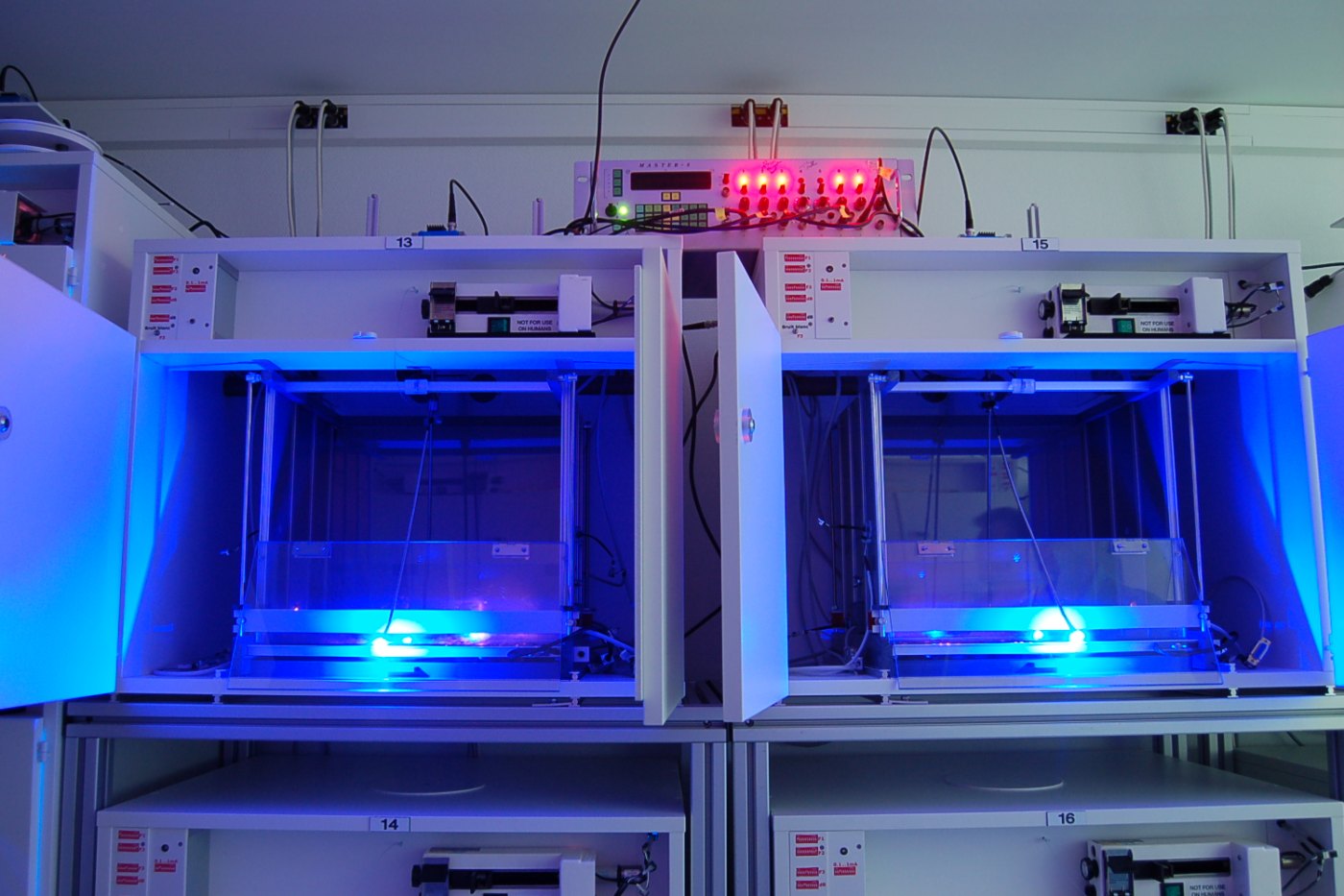
A mouse explores a small box, sniffing the corners. A researcher presses a button, a hair-thin glass fibre in the animal's head glows blue, and the mouse stops still, as though it has remembered something frightening. Nothing in the box has changed. What changed is a few thousand nerve cells deep inside its brain, switched on by a flash of light. This is optogenetics, and on 5 October 2026 it brought Karl Deisseroth, Peter Hegemann and Georg Nagel the Nobel Prize in Physiology or Medicine.
A wish written down in 1979
The human brain holds tens of billions of nerve cells of hundreds of different types, packed together and intertwined. For most of the twentieth century, researchers studied it with blunt instruments. An electrode could stimulate a patch of tissue, but it excited every cell nearby. Drugs acted over minutes or hours, far more slowly than thought itself. Injuring a region showed what was lost, but not which cells had been doing the work.
In 1979 Francis Crick, who with James Watson had described the double helix of DNA, set out what neuroscience was missing: a way to control one kind of cell in the brain and leave all the others untouched. Later he suggested that light might be the tool, because it is fast and can be aimed. The obvious problem was that nerve cells, unlike the light-detecting cells of the eye, pay no attention to light at all.
A gift from a green alga
The solution came from a pond. Chlamydomonas reinhardtii is a single-celled green alga that swims towards light, steered by a tiny orange eyespot. In the 1990s Peter Hegemann, then at the Max Planck Institute of Biochemistry near Munich, noticed that the alga answers light remarkably quickly, within about half a millisecond. In the human eye the same job takes at least 10 milliseconds, because it runs through a chain of several proteins. Hegemann suspected that in the alga a single protein does everything: catches the light and opens a pore in the cell membrane.
Colleagues were sceptical, and the fragile algal proteins were hard to study. The breakthrough came when Japanese researchers published the alga's genes. Hegemann found two promising candidates and sent them to Georg Nagel, a membrane specialist at the Max Planck Institute of Biophysics in Frankfurt. Nagel injected the genes into frog egg cells, which produced the unknown proteins, and measured what they did. Hegemann had been right. In 2002 and 2003 the team described channelrhodopsin-1 and channelrhodopsin-2: ion channels, meaning gates in the cell membrane that let electrically charged particles through, which open when blue light strikes them. Put the gene into almost any cell, the researchers showed, and that cell becomes sensitive to light.
Into a living brain
Other laboratories were chasing the same goal by other routes. Gero Miesenböck, then in New York, made cultured neurons respond to light in 2002 using light-sensing proteins taken from the fruit fly. That system worked, but it was slow and needed several genes. Channelrhodopsin-2 was a single gene, and it was fast.
Karl Deisseroth, a psychiatrist and bioengineer at Stanford University, had been looking for exactly such a switch. Treating patients with depression and schizophrenia had convinced him that progress required knowing what specific circuits actually do. He asked Nagel for the DNA, put it into rat nerve cells growing in a dish, and found that they tolerated the foreign protein well. In 2005 Deisseroth, Ed Boyden, Feng Zhang, Nagel and Ernst Bamberg reported that a pulse of blue light made these neurons fire an electrical spike, reliably and within milliseconds. The method was named optogenetics in 2006. In 2007 Deisseroth's group delivered the gene to the motor cortex of living mice, threaded a thin optical fibre through a small hole in the skull, and moved the animals' whiskers with light.
Optogenetics joins two technologies. The first is genetic. The gene for a light-sensitive protein, an opsin, is packaged into a harmless virus, usually an adeno-associated virus, and injected into a chosen brain region. Genetic address labels ensure that only one cell type, dopamine neurons for example, actually makes the protein.
The second is optical. A fine fibre, or in some experiments a light-emitting diode, carries light to those cells. When the light comes on, only the modified cells react; their neighbours stay as they were. Because opsins respond within milliseconds, researchers can imitate the brain's own rhythms. A growing toolkit now offers switches in several colours, including proteins such as halorhodopsin that silence a neuron rather than exciting it. The crucial gain is logical: instead of asking which cells are busy while an animal does something, scientists can ask what happens if these particular cells are switched on, or off, right now. That is the difference between a correlation and a cause.
What the light has shown
- Memory. In 2012 Susumu Tonegawa's laboratory at MIT labelled the hippocampal neurons that were active while mice learned to fear a particular box. Switching those same cells on with light in a different, safe box made the animals freeze: the memory had been recalled artificially. In 2013 Steve Ramirez, Xu Liu and Tonegawa went further and planted a fear memory of an event that had never happened.
- Emotion and motivation. Light-controlled experiments have mapped circuits for fear and anxiety in the amygdala, and reward circuits built on dopamine neurons, showing that activating them can drive an animal to work for the stimulation.
- Movement. In mouse models of Parkinson's disease, activating or silencing particular pathways in the basal ganglia changed the symptoms, helping to settle long arguments about how these circuits are organised.
- Beyond the brain. The same approach has been used in the heart, the gut and the sensory nerves, and has identified cells that govern thirst, appetite and sleep.
The first steps towards patients
In 2021 a team led by Jose-Alain Sahel of the University of Pittsburgh and the Sorbonne, with Botond Roska in Basel, reported in Nature Medicine the first clear clinical result. A 58-year-old man had been almost completely blind for decades because of retinitis pigmentosa, an inherited disease that destroys the light-sensitive cells of the retina. Researchers delivered the gene for a channelrhodopsin-type protein into surviving cells of one retina, and gave him goggles that convert the visual scene into pulses of amber light. Months later, he could locate, count and touch objects on a table in front of him, something he had not been able to do before. This is a single patient in an ongoing trial, not an approved treatment, and the vision regained was coarse rather than normal. Several companies are now testing similar therapies, and groups in Germany are developing optical cochlear implants that might stimulate the hearing nerve more precisely than electrical ones.
What it cannot do yet
Optogenetics remains, for now, overwhelmingly a laboratory method. It requires putting a foreign gene into cells, which for the brain means gene therapy with all its safety questions. Visible light is absorbed and scattered by tissue, so it reaches only a few millimetres and deep structures still need an implanted fibre. The eye is the exception that made the 2021 trial possible: it is accessible, transparent and relatively protected from the immune system. Researchers are working on opsins that answer to red or infrared light, which penetrates further, on ultrasound and magnetic alternatives, and on gentler ways to deliver genes.
Even if no one is ever treated for depression with a fibre-optic cable, the knowledge gathered with light is already changing medicine. Knowing which cells in a circuit produce a symptom tells drug developers what to aim at, and electrical stimulators where to go. A protein that evolved so that a pond alga could find sunlight has become one of the most precise instruments ever pointed at the brain: a useful reminder of why curiosity-driven biology is worth funding.
Sources: NobelPrize.org: The Nobel Prize in Physiology or Medicine 2026, popular information; Nature Neuroscience (2005), Boyden, Zhang, Bamberg, Nagel & Deisseroth: Millisecond-timescale, genetically targeted optical control of neural activity; PNAS (2003), Nagel, Szellas, Hegemann et al.: Channelrhodopsin-2, a directly light-gated cation-selective membrane channel; Nature (2012), Liu, Ramirez, Tonegawa et al.: Optogenetic stimulation of a hippocampal engram activates fear memory recall; Nature Medicine (2021), Sahel et al.: Partial recovery of visual function in a blind patient after optogenetic therapy. Image: Bd008 / Wikimedia Commons (CC BY-SA 4.0).

Comments
Please login to leave a comment Sign in