

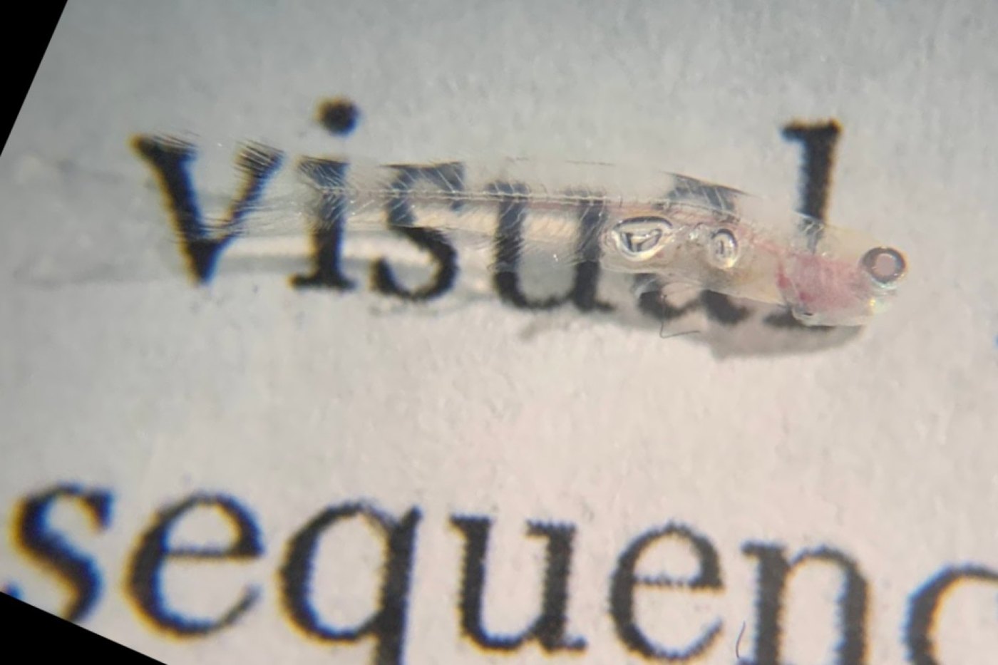
Look down on Danionella cerebrum and you can see its brain through the top of its head. This transparent fish, barely a centimetre long, has now given biologists something new: a map of the cell types and gene activity across an entire adult vertebrate body. Researchers profiled more than 600,000 cells drawn from across the fish, from brain to fins. The result is a reference for how cells in different tissues relate to one another, and how a change in one part of the body might ripple through the rest. The study was published in Science on 29 October.
Why size mattered
Cell atlases have grown steadily more detailed, but covering a whole adult vertebrate has been out of reach for a simple reason: most vertebrates are big. Cataloguing every kind of cell in a large animal takes a great deal of technical effort and money. Danionella, formally described as a species only in 2021, is small enough to make the job feasible. It is also a close relative of the zebrafish, so researchers can borrow genetic tools from one of biology's most popular lab animals.
The project was led by Kutay Deniz Atabay, then a postdoc in the lab of Peter Reddien, a Whitehead Institute member, MIT biology professor and Howard Hughes Medical Institute investigator. Collaborators at Charité–Universitätsmedizin Berlin also took part. Atabay first became interested in the fish in early 2019, when he was looking for a small, see-through animal in which he could watch the nervous system regenerate. Reddien's lab had already built a whole-animal atlas for planarians, tiny flatworms famous for regrowing lost body parts. Together, Reddien says, the two of them wondered: “Would it be possible to get a cell-type transcriptome atlas for a complete vertebrate?” As he puts it, “Here's an animal where we'd have a chance.”
Reading every cell's to-do list
The atlas is built on transcriptomes. A transcriptome is the full set of RNA molecules a cell has copied from its DNA at a given moment, which shows which genes the cell is actually using. The team applied single-cell RNA sequencing, which reads this activity in individual cells, to samples from the brain, spinal cord, eyes, internal organs, fins, head and trunk. They then used spatial techniques to place each cell type back where it sits in the tissue. The resulting map was thorough enough to pick up even very rare kinds of cells.
“Once you have a spatially resolved atlas, you know what cell types exist, but you also know the neighbors of those cell types in this context,” Atabay says.
Neurons grouped by neighbourhood, cells that remember their place
Viewing the whole animal at once revealed patterns that are hard to spot one tissue at a time. Neurons from the same brain region often resembled each other in gene activity more closely than neurons that used the same neurotransmitter (the chemical messenger nerve cells release) but lived in different regions. Comparable findings have been reported in other vertebrates, mammals among them. This suggests that where a neuron sits and how it developed may be a key organizing principle of vertebrate nervous systems.
The atlas also hinted that adult cells keep a molecular memory of their place in the body. Fibroblasts, connective-tissue cells, kept switching on region-specific genes best known for laying out the body plan in embryos. For instance, cells in the fish's pectoral and pelvic fins still used genetic programs that, in other vertebrates, are tied to the development of front and hind limbs. What these lingering signals do in the adult fish is still unknown. The researchers suggest they may help tissues maintain or rebuild the right structures, much as positional cues guide regeneration in planarians.
Watching a brain repair itself
The fish turns out to regenerate widely, including in its brain and fins. Using the atlas as a reference, the team damaged the telencephalon, part of the forebrain, and watched the lost neurons return. They traced a sequence that began with a wound and immune response, moved to neural stem-cell activity, and ended with new neurons being made and maturing.
An open resource
The atlas is freely available through an interactive tool called DigiFish. It offers a starting point for studying how the animal's cells respond everywhere in the body to injury, ageing, drugs and other influences. Because the fish is transparent, researchers can pick out particular cells or genes, manipulate them, and follow what happens in the living animal.
“To have the transcriptome of every cell type seems like a big turning point in how one would investigate the biology of an organism,” Reddien says.
Sources: MIT News | Massachusetts Institute of Technology; Science (original study). Image: Kutay Deniz Atabay / Whitehead Institute.


Comments
Please login to leave a comment Sign in