

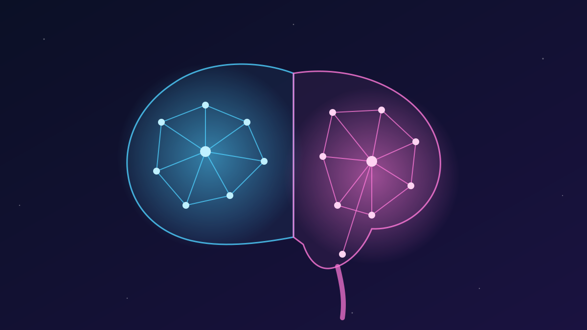
For decades, textbooks have described the brain as one organ that grows from a single pool of early cells, which gradually divides into the forebrain, midbrain and hindbrain. A new study led by Stanford Medicine and published on September 18, 2026 in Nature Neuroscience overturns that picture. According to the researchers, the brain is really two neural systems with separate origins that grow side by side and join up during development.
Two origins from the earliest stage
The team, led by senior author Kyle Loh with co-first authors Carolyn Dundes and Rayyan Jokhai, studied mouse embryos during gastrulation, the stage at which the body plan is first laid out. They found two distinct groups of brain progenitor cells. Cells that switch on the gene Otx2 give rise to the forebrain and midbrain, the regions behind language, reasoning and other higher functions. Cells that switch on Gbx2 form the hindbrain, the brain stem region that runs breathing, heartbeat and the muscles used to swallow and speak.
The two populations never overlap. The hindbrain does not branch off the forebrain lineage: it develops along its own parallel track from the start.
Locked in by chromatin
The researchers also compared chromatin, the packaging that controls which genes a cell can reach. The front (anterior) and back (posterior) neural tissue turned out to be packaged in fundamentally different ways. These differences appear to lock each progenitor into its fate, so one type cannot simply be converted into the other.
Hindbrain neurons in a dish, for the first time
This explains why labs have long struggled to grow hindbrain neurons. Earlier protocols probably tried to push forebrain-type progenitors into becoming hindbrain cells, which the study shows is not possible. Using the right starting population, the team turned human pluripotent stem cells into hindbrain motor neurons. The cells fired action potentials and carried the molecular markers of the brain stem segments that control facial muscles, speech and swallowing.
A pattern hundreds of millions of years old
The same two-origin plan appears in chickens, zebrafish and acorn worms, which means it has been conserved for more than 550 million years. Jellyfish, whose lineage split from ours roughly 600–700 million years ago, have two separate nervous systems at opposite ends of the body. The authors suggest that evolution may have taken two existing neural systems and brought them together into what we now call one brain.
Why it matters for patients
Brain stem neurons are among the first to fail in spinal muscular atrophy (SMA), the leading genetic cause of death in infants under one year old, and in amyotrophic lateral sclerosis (ALS), which is usually diagnosed between ages 40 and 70. As these neurons stop working, patients lose the ability to swallow and eventually to breathe. Living brain stem tissue cannot be taken from patients, so lab-grown hindbrain neurons give researchers a new way to study how these diseases develop. The hindbrain also contains circuits that regulate hunger, which are relevant to weight-loss drugs such as semaglutide.
Next, the team plans to trace where the spinal cord comes from and to study exactly how SMA and ALS damage hindbrain neurons. Their long-term goal is regenerative therapy for these conditions.
Source: Stanford Medicine news, September 2026; Nature Neuroscience.


Comments
Please login to leave a comment Sign in